DEVICE FOR CELL CULTURE AND ANALYSIS
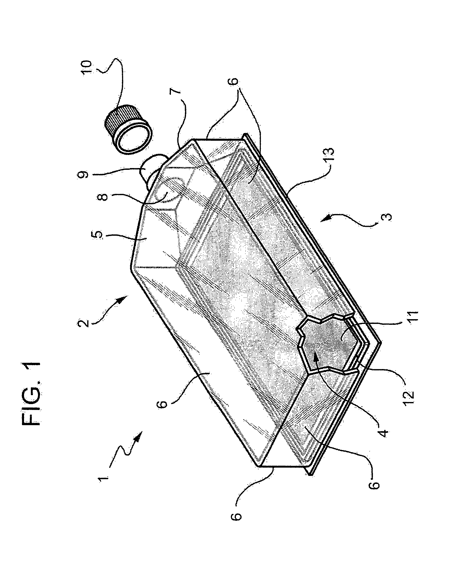
The present invention relates to a device for cell culture and analysis. It is known to use flasks for cell culture to allow cell growth in vitro. In particular, these flasks have a substantially parallelepipedal shape and are provided, on a face having a smaller surface with a tubular neck defining an opening adapted to be sealingly closed by a cap. The flask internally defines a parallelepipedal culture chamber. A culture medium is placed in the chamber by introduction through the neck; the cells are then seeded on this culture medium. In particular, the culture medium is normally placed on an inner face of the culture chamber that has a larger surface. To promote cell adhesion, the inner face of the culture chamber is treated, by means of known methods, to produce an electrostatically positively charged or negatively charged surface. After seeding the cells, the sealed flask is maintained in a controlled temperature environment to promote cell growth. In many cases, once a cell population is obtained, the presence of a specific group of proteins must be determined on the cell in order to characterise the cell type or determine the biological effect thereof. Protein expression is nowadays normally tested by immunofluorescence or immunohistochemistry. However, these methods are not suitable for the screening of many proteins in a high number of samples (high-throughput screening, HTP) both for the large amount of cells required, and due to the time- and material-consuming and expensive procedures. For example, up to now, the number of minimum starting cells for each single support (glass or flask) is about 50.000 units; the markers that may be characterised on the same support are normally less than 5 and the times are long, as the protocols are not automated. The need is therefore felt in the field for a device that allows to apply immunohistochemistry and immunofluorescence techniques even to high-throughput screening. In particular, the need is felt in the field to provide a device that makes lab tests simpler for characterising primary cell lines and stem cells after protocols for differentiation of neoplastic populations, of immunophenotypic alterations, as well as for identifying new drugs and evaluating their biological effect. It is the object of the present invention to provide a cell growth device which allows to satisfy in a simple and cost-effective manner one of the above said needs. This object is achieved by the present invention, as it relates to a cell culture and cell analysis device according to claim 1 and a method for cell analysis according to claim 11. A preferred embodiment is hereinafter disclosed for a better understanding of the present invention, by mere way of non-limitative example and with reference to the accompanying drawings, in which: Flask 1 comprises a first containing element 2 and a second containing element 3 sealingly couplable to one another to define a culture chamber 4 (in First element 2 comprises a bottom wall 5 having a substantially rectangular flat shape integral with side walls 6 which extend along peripheral edges of the bottom wall 5 and are transversal to the bottom wall 5. In the figures, side walls 6 are shown arranged perpendicularly with respect to bottom wall 5. In particular, a cylindrical tubular appendix 9 extends integrally from a central portion 7 of a side wall 6 having, in the embodiment shown, a smaller surface. Tubular appendix 9 extends outwards from device 1 and defines a circular opening 8 to allow the communication with the outside of culture chamber 4, for example to introduce/replace the culture medium. Tubular appendix 9 may be closed by removable sealing means 10, for example a cap which is screwed/unscrewed on the end portion of tubular appendix 9 which for this purpose is provided with helicoidal ribs. Second element 3 has a substantially flat rectangular configuration and is provided with peripheral edges 13 adapted to sealingly couple with corresponding peripheral edges 14 of side walls 6. In use, second element 3 forms a wall of flask 1 arranged spatially in a facing position opposite to bottom wall 5. Second rectangular flat element 3 may have dimensions substantially corresponding to that of bottom wall 5 as shown in Advantageously, the embodiment shown in First element 2 and second element 3 can be sealingly coupled by means of snap coupling means 12, for example a peripheral groove made on the inner surface of second element 3 adapted to house at least the end portion of side walls 6 of first element 2, or alternatively a peripheral projection on the inner surface of second element 3 that abuts with side walls 6 of first element 2. Alternatively the sealing coupling between first element 2 and second element 3 can be made by ultrasound welding and/or glues. Coupling means 12 are such as to maintain the impermeability of the flask to the outside environment in use, although they can be manually forced to allow the operation of first element 2 and of second element 3 and therefore the opening of flask 1 making the rinsing and possibly a second use easier. First element 2 and second element 3 are made of a rigid and transparent material such as for example polystyrene, polycarbonate or glass. Flask 1 further comprises a flexible film 11 housable within culture chamber 4 and available on the inner surface of second rectangular flat element 3 or alternatively on the inner surface of bottom wall 5 of first element 2. In the embodiment of the figures, film 11 has a rectangular perimeter. This flexible film 11 is treated by means of known methods to obtain an electrostatically positive or negative charged surface to provide cell growth. It is made of a transparent material, preferably selected from the group consisting of polycarbonate, polystyrene and polyethylene, to allow its use for example in microscope analysis. Furthermore, the flexibility of the material advantageously allows to roll and fold film 11 for subsequent immunofluorescence or immunohistochemistry analysis. As shown in In use, flexible film 11 is fixed on second element 3, for example by means of an adhesive. Subsequently, second element 3 is coupled to first element 2 so as to arrange flexible film 11 within chamber 4 and allow the laying and cell growth on flexible film 11. Alternatively, flexible film 11 can be fixed interposing it between first element 2 and second element 3, coupling means 12 of which retain a peripheral portion of flexible film 11. Advantageously, after the cell culture cycle has been completed or the fresh sample has been laid, flexible film 11 carrying the cells is easily and rapidly removed from culture chamber 4 as the uncoupling of elements 2, 3 is simple and fast and uncoupled second element 3 forms a support for flexible film 11. Thereby, flexible film 11 can be subjected directly to biological analyses such as immunohistochemistry and immunofluorescence. Flexible film 11 is also compatible with other standard hybridisation techniques such as FISH and in situ hybridisation. Finally, the cells on flexible film 11 can be fixed with formaldehyde or paraformaldehyde, can be included in paraffin and analysed directly using HTS systems to identify the expression profile of the cells. In Similarly to what has been disclosed for Finally, it is clear that modifications and variants not departing from the scope of protection of the independent claims can be made to the disclosed and shown system. The present invention relates to a device for cell culture and analysis, comprising a first containing element, a second containing element sealingly couplable to one another to define a culture chamber and a flexible film housable in said culture chamber and available alternatively on one of said first element or second element. The flexible film can be made of a transparent material selected from the group consisting of polycarbonate, polystyrene and polyethylene. A method for cell analysis is also provided. 1. A device (1) for cell culture and analysis, comprising a first containing element (2), a second containing element (3) sealingly couplable to one another to define a culture chamber (4) and a flexible film (11) housable in said culture chamber (4) and available alternatively on one of said first element (2) or second element (3). 2. The device according to 3. The device according to 4. The device according to 5. The device according to said second element (3) comprising a substantially flat wall provided with peripheral edges (13) adapted to sealingly couple with corresponding peripheral edges (14) of said side walls (6). 6. The device according to 7. The device according to 8. The device according to 9. The device according to 10. The device according to any of the preceding claims, wherein said first element (2) and said second element (3) are sealingly coupled by snap coupling means (12). 11. A method for cell analysis comprising the steps of:
a) culturing the cells to be analysed on a flexible film (11); b) winding said flexible film (11) by means of winding means (16) to obtain a cylinder (17); c) providing a support (18) provided with at least one cavity (19) having dimensions substantially corresponding to those of the cylinder (17); d) inserting at least one of said cylinders (17) in a respective cavity (19); e) sectioning said support (18) along a direction transversal to the axis of said cavity (19) so as to obtain at least one section (20) of said support (18) incorporating a section of said cylinder (17); f) subjecting said section (20) to said cell analysis. 12. The method according to 13. The method according to TECHNICAL FIELD
BACKGROUND ART
DISCLOSURE OF INVENTION
BRIEF DESCRIPTION OF THE DRAWINGS
BEST MODE FOR CARRYING OUT THE INVENTION